


Varicose veins in the lower extremities are characterized by expansion of the superficial veins in the legs, which accompanies the violation of blood flow in the legs and valve failure. As a result, the veins increase in length and diameter, acquire a serpentine, cylindrical or saccular aspect, although there is also a mixed manifestation of the listed deformities.
Characteristics of the venous system
The emergence and development of varicose veins is directly related to the venous system of the legs, which consists of:
- saphenous veins: small and large;
- veins located deep (in the lower leg and thigh);
- perforating veins, which are the connecting link of the two anterior systems.
Typically, 90% of blood is transported to the lower extremities through deep veins and the remaining 10% through superficial veins. When it returns to the side of the heart, this mechanism is supported by valves in the walls of the veins. When the next bit of blood arrives, they knock to prevent its upward movement under the influence of gravitational force. Muscle contractions push blood to the heart, allowing normal blood flow.
With a long stay of a person in an upright position, blood stagnation can occur, which increases the pressure in the veins and causes an increase in their diameter. This process causes the incomplete closure of the valve leaflets, in which the blood flow is disturbed with its reverse flow from the heart - reflux.
The deep vein valves are more likely to be affected as they carry the greatest amount of blood and therefore bear the maximum load. To reduce the high pressure in them, part of the blood is transported through perforated veins to the superficial ones, which were not originally intended for a large volume. This load on the walls of the veins leads to their expansion and the formation of varicose veins.
At the same time, blood enters the deep veins without stopping, but due to the violation of their functions and the normal activity of the valve leaflets of the perforated veins, the blood is redistributed to the superficial vessels. As a result, chronic varicose veins appear, which over time are accompanied by painful sensations, edema, and trophic ulcers.
Causes of disease
Previously, one of the main causes of varicose veins was called the hereditary factor, but today this theory has been refuted. Of course, it is possible to track the frequent manifestations of the disease in some families, but this is more likely due to the peculiarities of life that are transmitted to the family: food culture, passive rest, sedentary work and so on.
The development of varicose veins is based on the presence of reflux in the venous system, when blood circulates through the veins in the opposite direction. The additional transport of blood from the veins located deep to the superficial veins is possible due to the congenital or acquired degenerative pathology of the valve apparatus. This causes the superficial vessels to overfill with blood and distend them when venous nodules form.
One of the fundamental reasons for the development of varicose veins is considered an unhealthy diet, which in some cases leads to obesity. These people move little, eat mostly highly processed foods, and the proportion of vegetable fiber in the diet is minimized. After all, they are the ones who strive to strengthen the walls of veins and blood vessels and prevent prolonged chronic constipation, which greatly increase intra-abdominal pressure and, therefore, cause varicose veins. It is observed that an increase in body weight of more than 20% increases the risk of disease fivefold.
The main provocative factor for women is pregnancy, while the risk of varicose veins increases with each subsequent pregnancy. Great weight gain and an enlarged uterus put a lot of stress on the stagnant legs. This situation is aggravated by the constant increase in intra-abdominal pressure and by the action of the hormone progesterone, which affects the state of elastic fibers in the walls of blood vessels.
Other factors that cause varicose veins in the lower extremities include:
- a sedentary lifestyle, standing during the day (eg hairdressers), long flights or long journeys. All of this leads to stagnation processes in the lower extremities, when blood accumulates in the superficial veins and is poorly transported to the heart;
- sometimes increases the risk of developing varicose veins for women, wearing tight and uncomfortable shoes, especially models with high heels;
- corsets and tight underwear compress the inguinal veins and increase intra-abdominal pressure, which is a direct prerequisite for varicose veins;
- high pressure;
- smoking, which indirectly leads to a thinning of blood vessel walls.
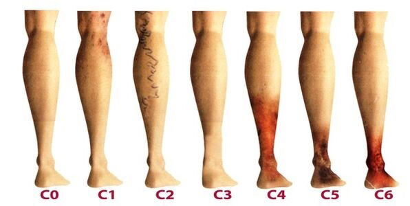
Disease classification
Lower limb varicose veins are classified according to the prevalence of venous lesions, their location, as well as the presence of pathological reflux, which is characterized by compromised blood flow. There are 4 forms of varicose veins:
- intracutaneous and subcutaneous (segmental) varicose veins, in which there is no pathological outflow of venous blood;
- segmental varicose veins, when reflux occurs through perforating or superficial veins;
- a common form of varicose veins, in which reflux occurs through perforating and superficial veins at the same time;
- varicose veins are characterized by reflux in the deep veins.
After varicose veins in the lower extremities become chronic, phlebology considers its three degrees:
The last degree is the most difficult to treat, as it requires preliminary removal of inflammation and healing of the skin tissues.
Stages and symptoms
The disease develops very slowly, sometimes more than a dozen years pass, until the symptoms that appear will force the patient to consult a phlebologist. In the early stages of varicose veins, its manifestations are often attributed to fatigue, age, or other reasons. To fully consider the symptoms of the disease, its manifestations are classified according to the stages of the varicose veins:
Diagnosis
An external examination of the lower extremities in the vertical and horizontal positions of the body, palpation of the veins and preliminary assessment of the stage of the disease are performed. The patient is referred for a general blood test, which allows a more in-depth study of the disease picture:
- at the platelet level, a predisposition to thrombosis will be reflected;
- the hemoglobin level as well as the number of red blood cells indicate the degree of clotting of the blood;
- by increasing the level of white blood cells, one can judge inflammation, which helps to diagnose thrombophlebitis more quickly.
Be sure to examine the venous system in the legs, for which there are many methods:
- ultrasonographic dopplerography - USDG;
- phlebography;
- CT Phlebography;
- duplex angioscanning - USAS;
- phleboscintiography;
- photopolymography;
- phlebomanometry and the like.
In practice, patients are more often given USAS and USG as they help to thoroughly study the venous system in the legs and identify degenerative areas. The other methods can be additionally prescribed if the ultrasound exam does not provide a complete picture of the disease picture. Some of these methods can present complications such as venous thrombosis, catheter perforation of the vessel wall and allergy to contrast agents. Consider the techniques most commonly practiced in phlebology:
After extensive diagnoses, the physician prepares the patient's phlebocard, which makes it possible to determine the injured segments of the venous system, their degree and length. After that, a suitable treatment is selected.
Treatment
It is performed comprehensively and determined based on symptoms, degree of disease progression, and study results. In the early stages, conservative therapy is prescribed, which consists of:
- anti-protectives and phlebotonics;
- anticoagulants;
- disintegrators
- topical preparations (ointments, gels);
- anti-inflammatory drugs.
In addition, patients are advised to perform contrasting five-minute shower procedures every night, alternating between hot and cold water. These manipulations improve blood flow and tone blood vessels.
It is important, at the beginning of treatment, to identify the triggering factor of the disease to effectively influence it. And patients who are at risk should visit a phlebologist every 2 years for a preventive exam and have an ultrasound examination of the leg veins.
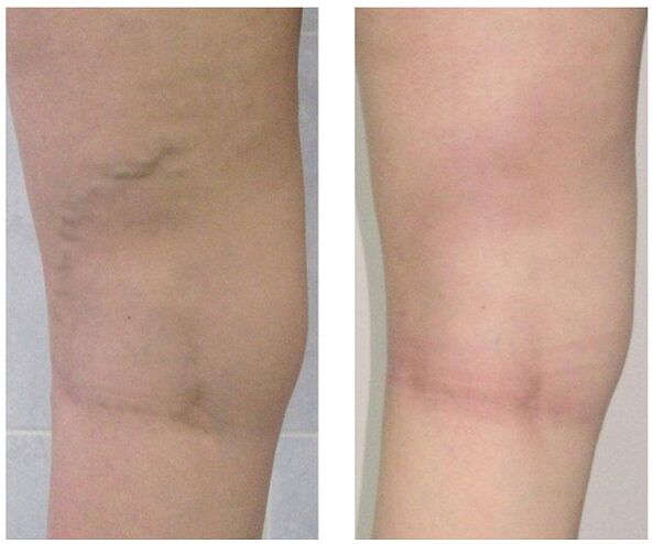
When conservative treatment does not work or varicose veins are seen in an advanced stage, surgical intervention is used. Today, varicose veins can be completely cured thanks to the following methods:
Prevention
Preventive measures can be primary, aimed at preventing the development of varicose veins, and secondary, when they are necessary to reduce the risk of recurrence after surgery or to prevent a worsening of the disease course. Helpful hints:
- lead an active lifestyle without overloading your legs: swimming, walking, cycling;
- pay attention to your weight;
- keep both legs raised more often;
- do not wear tight underwear and heels over 4 centimeters;
- use orthopedic insoles;
- take a contrast bath;
- do preventive leg exercises for five minutes daily;
- wear compression socks for long walks.
If you notice the slightest suspicion of varicose veins - prominent nodules in the legs, swelling, heaviness, don't postpone your visit to the phlebologist. In fact, over time, this insidious disease can lead to many complications, including thrombophlebitis and thrombosis.